A team spanning UCL institutions has identified markers picked up via eye scans that indicate the presence of Parkinson’s disease in patients on average seven years before clinical presentation.
It is the first time anyone has shown these findings several years before diagnosis, and these results were made possible by the largest study to date on retinal imaging in Parkinson’s disease.
Co-authors of the research Prof Anthony Schapira and Dr Rimona Weil are supported by the BRC at UCLH and the research is led by Siegfried Wagner and Pearse Keane of Moorfields Eye Hospital and UCL Institute of Ophthalmology.
The study, published in Neurology, the medical journal of the American Academy of Neurology, identified markers of Parkinson’s in eye scans with the help of artificial intelligence (AI). Its analysis of the AlzEye dataset was repeated using the wider UK Biobank database (healthy volunteers), which replicated the discoveries.
The use of these two large, powerful datasets enabled the team to identify these subtle markers, even though Parkinson’s disease has a relatively low prevalence (0.1-0.2% of the population). Generation of the AlzEye dataset was enabled by INSIGHT, the world's largest database of retinal images and associated clinical data.
The use of data from eye scans has previously revealed signs of other neurodegenerative conditions, including Alzheimer’s, multiple sclerosis and, most recently, schizophrenia, in an emerging and exciting field of research referred to as “oculomics”.
Eye scans and eye data have also been able to reveal a propensity to high blood pressure; cardiovascular disease including strokes; and diabetes.
Doctors have known for a long time that the eye can act as a ‘window’ to the rest of the body, giving a direct insight into many aspects of our health. High-resolution images of the retina are now a routine part of eye care – in particular, a type of 3D scan known as ‘optical coherence tomography’ (OCT), which is widely used in eye clinics and high-street opticians. In less than a minute, an OCT scan produces a cross-section of the retina (the back of the eye) in incredible detail – down to a thousandth of a millimetre.
These images are extremely useful for monitoring eye health, but their value goes much further, as a scan of the retina is the only non-intrusive way to view layers of cells below the skin’s surface. In recent years, researchers have started to use powerful computers to accurately analyse large numbers of OCTs and other eye images, in a fraction of the time it would take a human. Using a type of AI known as ‘machine learning’, computers are now able to uncover hidden information about the whole body from these images alone. Harnessing this new potential is what oculomics is about.
This work has involved collaboration between the NIHR BRCs at UCLH, Moorfields Eye Hospital, University Hospital Birmingham, Great Ormond Street Hospital, Oxford University Hospital, and the UCL Great Ormond Street Institute of Child Health and UCL Queen Square Institute of Neurology.
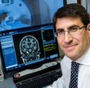
Photo: Dr Siegfried Wagner giving a patient with Parkinson's an OCT eye scan